
A hand (med./lat.: manus, pl. manūs) is a prehensile, multi-fingered extremity located at the end of an arm or forelimb of primates such as humans, chimpanzees, monkeys, and lemurs.

A few other vertebrates such as the koala (which has two opposable thumbs on each "hand" and fingerprints remarkably similar to human fingerprints) are often described as having either "hands" or "paws" on their front limbs.

Hands are the main structures for physically manipulating the environment, used for both gross motor skills (such as grasping a large object) and fine motor skills (such as picking up a small pebble).

The fingertips contain some of the densest areas of nerve endings on the body, are the richest source of tactile feedback, and have the greatest positioning capability of the body; thus the sense of touch is intimately associated with hands. Like other paired organs (eyes, feet, legs), each hand is dominantly controlled by the opposing brain hemisphere, so that handedness, or the preferred hand choice for single-handed activities such as writing with a pen, reflects individual brain functioning.

Some evolutionary anatomists use the term hand to refer to the appendage of digits on the forelimb more generally — for example, in the context of whether the three digits of the bird hand involved the same homologous loss of two digits as in the dinosaur hand.
 |
| Michael Jordan's Hand, |
The human hand has 27 bones, 14 of which are the phalanges (proximal, medial, and distal) of the fingers. The metacarpal is the bone that connects the fingers and the wrist. Each human hand has 5 metacarpals.

Definition
the terminal, prehensile part of the arm in humans and higher primates, consisting of the wrist, metacarpals, fingers, and thumb.
Human anatomy
The anatomy of the hand is complex, intricate, and fascinating. Its integrity is absolutely essential for our everyday functional living. Our hands may be affected by many disorders, most commonly traumatic injury. For any physician or therapist treating hand problems, the mastery of such anatomy is fundamental in order to provide the best quality of care.

Surface anatomy of the left hand. A is the dorsum of the left hand, and B is the palm of the left hand. Number 1 is the position of the extensor retinaculum, 2 is the position of the flexor retinaculum, 3 is the position of the head of the metacarpals, 4 is the ulnar artery, 5 is the radial artery, 6 is the level of the deep palmar arch, and 7 is the level of the superficial palmar arch.
A total of 27 bones constitute the basic skeleton of the wrist and hand. The hand is innervated by 3 nerves — the median, ulnar, and radial nerves — each of which has sensory and motor components. The muscles of the hand are divided into intrinsic and extrinsic groups.
Bones
As previously mentioned, a total of 27 bones constitute the basic skeleton of the wrist and hand. These are grouped into carpals, metacarpals, and phalanges.
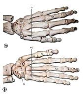
Bones of the left hand. A is the dorsal view, and B is the palmar view. Number 1 is the radius, 2 is the ulna, 3 is the scaphoid, 4 is the lunate, 5 is the triquetral, 6 is the pisiform, 7 is the trapezium, 8 is the trapezoid, 9 is the capitate, 10 is the hamate, 11 is the metacarpal bones, 12 is the proximal phalanx, 13 is the middle phalanx, and 14 is the distal phalanx.
The wrist is the most complex joint in the body. It is formed by 8 carpal bones grouped in 2 rows with very restricted motion between them. From radial to ulnar, the proximal row consists of the scaphoid, lunate, triquetrum, and pisiform bones. In the same direction, the distal row consists of the trapezium, trapezoid, capitate, and hamate bones.
All carpal bones participate in wrist function except for the pisiform, which is a sesamoid bone through which the flexor carpi ulnaris tendon passes. The scaphoid serves as link between each row; therefore, it is vulnerable to fractures. The distal row of carpal bones is strongly attached to the base of the second and third metacarpals, forming a fixed unit. All other structures (mobile units) move in relation to this stable unit. The flexor retinaculum, which attaches to the pisiform and hook of hamate ulnarly and to the scaphoid and trapezium radially, forms the roof of the carpal tunnel.
The hand contains 5 metacarpal bones. Each metacarpal is characterized as having a base, a shaft, a neck, and a head. The first metacarpal bone (thumb) is the shortest and most mobile. It articulates proximally with the trapezium. The other 4 metacarpals articulate with the trapezoid, capitate, and hamate at the base. Each metacarpal head articulates distally with the proximal phalanges of each digit.
The hand contains 14 phalanges. Each digit contains 3 phalanges (proximal, middle, and distal), except for the thumb, which only has 2 phalanges. To avoid confusion, each digit is referred to by its name (thumb, index, long, ring, and small) rather than by number.
Skin
The skin that covers the dorsum of the hand is greatly different from the skin that covers the palm. (See the image below.) The skin of the dorsum of the hand is thin and pliable. It is attached to the hand's skeleton only by loose areolar tissue, where lymphatics and veins course. This fact explains why edema of the hand is manifested predominantly at the dorsum. In addition, this loose attachment of skin makes the dorsum of the hand more vulnerable to skin avulsion injuries and also permits the creation of local flaps.
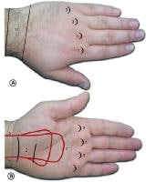 Surface anatomy of the left hand. A is the dorsum of the left hand, and B is the palm of the left hand. Number 1 is the position of the extensor retinaculum, 2 is the position of the flexor retinaculum, 3 is the position of the head of the metacarpals, 4 is the ulnar artery, 5 is the radial artery, 6 is the level of the deep palmar arch, and 7 is the level of the superficial palmar arch.
The skin of the palmar surface of the hand is unique, with characteristics for special function. The palmar skin is thick and glabrous and not as pliable as the dorsal skin. It is strongly attached to the underlying fascia by numerous vertical fibers. These features enhance skin stability for proper grasping function.
The skin is most firmly anchored to the deep structures at the palmar creases; this is of clinical importance when planning surgical incisions, to minimize skin contractures. In contrast to the dorsal skin, the blood supply to the palmar skin is through numerous small, vertical branches from the common digital vessels. Therefore, the elevation of palmar skin flaps is limited. Finally, the skin of the palmar surface of the hand contains a high concentration of sensory nerve organs essential to the hand's normal function.
Nails
Sexual dimorphism
Sexual dimorphism
The nails are specialized skin appendages derived from the epidermis. The nail bed has a germinal matrix, sterile matrix, and hyponychium. Ninety percent of the nail plate is produced by the germinal matrix, which approximately corresponds to the lunula (pale semicircle in proximal nail bed). This germinal matrix starts proximally at the base of the distal phalanx just distal to the insertion of the extensor tendon.
The sterile matrix is distal to the lunula; it is very vascular, which accounts for the pink color. The sterile matrix produces 10% of nail plate volume and adds squamous components, which make the nail stronger and adherent to the nail bed. The hyponychium is the distal part of the nail bed; its abundance of immune cells and adherence to the distal nail plate help the nail to resist infection.
The entire nail matrix is in intimate contact with the periosteum of the distal phalanx; therefore, it is vulnerable to injury when the latter is fractured.
The average length of an adult male hand is 189 mm, while the average length of an adult female hand is 172 mm. The average hand breadth for adult males and females is 84 and 74 mm respectively.
Disorders and diseases
- Polydactyly, more than the usual number of fingers or toes. Having more than the typical number of metacarpal bones may be caused by genetic disorders like Catel-Manzke syndrome.
- Syndactyly, fusion of fingers or toes.
- Hand infection
- Hand surgery
- Carpal tunnel syndrome
- Dupuytren's contracture
- Claw hand, paralysis of intrinsic muscles of hand.
- Scaphoid fracture
- Rolando fracture - a comminuted intra-articular fracture through the base of the first metacarpal bone
- Bennett's fracture - a fracture of the base of the first metacarpal bone which extends into the carpometacarpal (CMC) joint.
- Boxer's fracture - a fracture at the neck of a metacarpal
 |  | |
A gorilla, a large primate with small thumbs, and the hand skeleton of Ardipithecus ramidus, a small Pliocene primate with human-like thumbs
| ||
Fractures of the hand include:
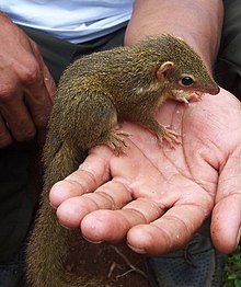
The prehensile hands and feet of primates evolved from the mobile hands of semi-arboreal tree shrews that lived about a100 million years ago. This development has been accompanied by important changes in the brain and the relocation of the eyes to the front of the face, together allowing the muscle control and stereoscopic vision necessary for controlled grasping. This grasping, also known as power grip, is supplemented by the precision grip between the thumb and the distal finger pads made possible by the opposable thumbs. Hominidae (great apes including humans) acquired an erect bipedal posture about 3 million years ago, which freed the hands from the task of locomotion and paved the way for the precision and range of motion in human hands. Functional analyses of the features unique to the hand of modern humans have shown that they are consistent with the stresses and requirements associated with the effective use of paleolithic stone tools. It is possible that the refinement of the bipedal posture in the earliest hominids evolved to facilitate the use of the trunk as leverage in accelerating the hand.
While the human hand has unique anatomical features, including a longer thumb and fingers that can be controlled individually to a higher degree, the hands of other primates are anatomically similar and the dexterity of the human hand can not be explained solely on anatomical factors. The neural machinery underlying hand movements is a major contributing factor; primates have evolved direct connections between neurons in cortical motor areas and spinal motoneurons, giving the cerebral cortex monosynaptic control over the motoneurons of the hand muscles; placing the hands "closer" to the brain. The recent evolution of the human hand is thus a direct result of the development of the central nervous system, and the hand, therefore, is a direct tool of our consciousness — the main source of differentiated tactile sensations — and a precise working organ enabling gestures — the expressions of our personalities.
There are nevertheless several primitive features left in the human hand, including pentadactyly (having five fingers), the hairless skin of the palm and fingers, and the os centrale found in human embryos, prosimians, and apes. Furthermore, the precursors of the intrinsic muscles of the hand are present in the earliest fishes, reflecting that the hand evolved from the pectoral fin and thus is much older than the arm in evolutionary terms.
The proportions of the human hand are plesiomorphic (shared by both ancestors and extant primate species); the elongated thumbs and short hands more closely resemble the hand proportions of Miocene apes than those of extant primates.Humans did not evolve from knuckle-walking apes, and chimpanzees and gorillas independently acquired elongated metacarpals as part of their adaptation to their modes of locomotion.Several of primitive hand features most likely present in the chimpanzee-human last common ancestor (CHLCA) and absent in modern humans are still present in the hands of Australopithecus, Paranthropus, and Homo floresiensis. This suggests that the derived changes in modern humans and Neanderthals did not evolve until 2.5 to 1.5 million years ago or after the appearance of the earliest Acheulianstone tools and that these changes are associated with tool-related tasks beyond those observed in other hominins.The thumbs of Ardipithecus ramidus, a CHLCA candidate, are robust like in humans, and thus a primitive trait, while the palms of other extant higher primates are elongated to the extent that some of the thumb's original function has been lost (most notably in highly arboreal primates such as the spider monkey). In humans, the big toe is thus more derived than the thumb.


0 comments