
A nail

or fingernail is a horn-like envelope covering the dorsal aspect of the terminal phalanges of fingers and toes in humans, most non-human primates, and a few other mammals.

Nails are similar to claws, which are found on numerous other animals.

Fingernails and toenails are made of a tough protein called keratin, as are animals' hooves and horns. The mammalian nail, claw, and hoof are all examples of unguis .


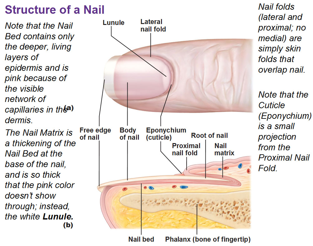
Parts of the nail
The matrix (synonyms: matrix unguis, keratogenous membrane, nail matrix, onychostroma) is the tissue (or germinal matrix) upon which the nail rests, the part of the nail bed that extends beneath the nail root and contains nerves, lymph and blood vessels.

The matrix is responsible for the production of the cells that become the nail plate.

The width and thickness of the nail plate is determined by the size, length, and thickness of the matrix, while the shape of the fingertip itself determines if the nail plate is flat, arched or hooked.

The matrix will continue to grow as long as it receives nutrition and remains in a healthy condition.

As new nail plate cells are incubated, they emerge from the matrix round and white to push older nail plate cells forward; and in this way yet older cells become compressed, flat, and translucent, making the pink colour of the capillaries in the nail bed below visible.

The lunula (occasionally called simply "the moon") is the visible part of the matrix, the whitish crescent-shaped base of the visible nail.

The lunula is largest in the thumb and often absent in the little finger.


The lunula is largest in the thumb and often absent in the little finger.

The nail bed is the skin beneath the nail plate.

Like all skin, it is composed of two types of tissues: the deeper dermis, the living tissue fixed to the bone which contains capillaries and glands,and the superficial epidermis, the layer just beneath the nail plate which moves forward with the plate.

The epidermis is attached to the dermis by tiny longitudinal "grooves" known as the matrix crests or crests of nail matrix (cristae matricis unguis).

During old age, the plate thins and these grooves are made evident in the structure.
.jpg)
The nail sinus (sinus unguis) is the deep furrow into which the nail root is inserted.


The nail root (radix unguis) is the part of nail situated in the nail sinus, i.e. the base of the nail embedded underneath the skin. It originates from the actively growing tissue below, the matrix.


The nail plate or body of nail (corpus unguis) is the actual nail, and like hair and skin, made of translucent keratin protein made of amino acids.

In the nail it forms a strong flexible material made of several layers of dead, flattened cells.

The plate appears pink because of the underlying capillaries.

Its (transversal) shape is determined by the form of the underlying bone. In common usage, the word nail often refers to this part only.


In the nail it forms a strong flexible material made of several layers of dead, flattened cells.

The plate appears pink because of the underlying capillaries.

Its (transversal) shape is determined by the form of the underlying bone. In common usage, the word nail often refers to this part only.

The free margin (margo liber) or distal edge is the anterior margin of the nail plate corresponding to the abrasive or cutting edge of the nail.

The hyponychium (informally known as the "quick") is the epithelium located beneath the nail plate at the junction between the free edge and the skin of the fingertip.

It forms a seal that protects the nail bed.

The onychodermal band is the seal between the nail plate and the hyponychium.

It is found just under the free edge, in that portion of the nail where the nail bed ends and can be recognized by its glassy, greyish colour (in fair-skinned people).
 It is not perceptible in some individuals while it is highly prominent on others.


The hyponychium (informally known as the "quick") is the epithelium located beneath the nail plate at the junction between the free edge and the skin of the fingertip.

It forms a seal that protects the nail bed.
The onychodermal band is the seal between the nail plate and the hyponychium.

It is found just under the free edge, in that portion of the nail where the nail bed ends and can be recognized by its glassy, greyish colour (in fair-skinned people).


The eponychium is the small band of epithelium that extends from the posterior nail wall onto the base of the nail.Often and erroneously[contradictory] called the "proximal fold" or "cuticle", the eponychium is the end of the proximal fold that folds back upon itself to shed an epidermal layer of skin onto the newly formed nail plate.

This layer of non-living, almost invisible skin is the cuticle that "rides out" on the surface of the nail plate. Together, the eponychium and the cuticle form a protective seal.

and is often removed during manicure, but the eponychium is living cells and should not be touched.

The perionyx is the projecting edge of the eponychium covering the proximal strip of the lunula.

This layer of non-living, almost invisible skin is the cuticle that "rides out" on the surface of the nail plate. Together, the eponychium and the cuticle form a protective seal.
and is often removed during manicure, but the eponychium is living cells and should not be touched.

The perionyx is the projecting edge of the eponychium covering the proximal strip of the lunula.

The nail wall (vallum unguis) is the cutaneous fold overlapping the sides and proximal end of the nail.

The lateral margin (margo lateralis) is lying beneath the nail wall on the sides of the nail and the nail groove or fold (sulcus matricis unguis) are the cutaneous slits into which the lateral margins are embedded.


The lateral margin (margo lateralis) is lying beneath the nail wall on the sides of the nail and the nail groove or fold (sulcus matricis unguis) are the cutaneous slits into which the lateral margins are embedded.

The paronychium is the border tissue around the nail and paronychia is an infection in this area.


Nail Polish Know-How
How to Repair a Torn Fingernail
THE EASY WAY: Nail Fungus Cures


0 comments